Back Of Skull Anatomy : Human skull, 3/4 back and top view. | Human skull, Skull anatomy, Skulls drawing. Human skull from the front. The base of the skull (or skull base) forms the floor of the cranial cavity and separates the brain from the structures of the neck and face. Atlas of human skeletal anatomy. Cranium) is the skeleton of the head composed of 22 separate bones joined together primarily by sutures. These joints fuse together in adulthood.
The major sutures are the coronal suture, sagittal suture, lambdoid suture and squamosal sutures. So, the human skull consists of 23 bones. A cartilaginous mould begins to grow this is why raising your eyebrows can create the appearance that the back of the head is moving. In order to be light, the skull is made up by flat and irregular bones, and has hollow spaces called the sinuses. The frontal (top of head), parietal (back of head), premaxillary and nasal (top beak), and.
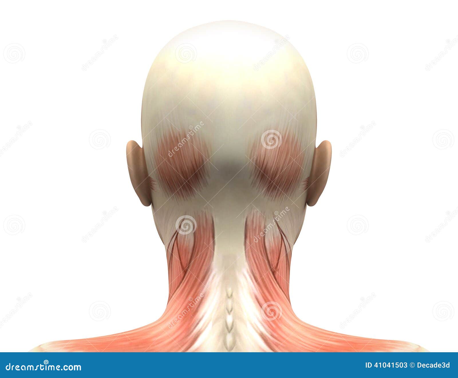
Female Head Muscles Anatomy - Back View Stock Illustration - Illustration: 41041503 from thumbs.dreamstime.com Cranium) is the skeleton of the head composed of 22 separate bones joined together primarily by sutures. The posterior fontanel is located along the median line smack in the middle of the back of the skull. Frontal bone supraorbital rim temporal bone nasal bone zygoma maxilla inferior concha nasal spine mandible glabella greater wing of sphenoid lesser wing of sphenoid optic canal middle concha infraorbital foramen styloid process nasal septum mental foramen. Human anatomy for muscle, reproductive, and skeleton. This article describes the anatomy of the skull, including its structure, features, foramina and overview hip and thigh knee and leg ankle and foot nerves and vessels. It was then cleaned, adapted and polypainted this model is part of a comparison with the skull of a human. Their number and location vary. It is comprised of many bones, formed by intramembranous ossification, which are joined together by sutures (fibrous joints).
Foramina inside the body of humans and other animals.
The major sutures are the coronal suture, sagittal suture, lambdoid suture and squamosal sutures. A thorough description is beyond the. The two fontanels located on the sides of the skull are mirror. The skull includes the upper jaw and the cranium. The posterior fontanel is located along the median line smack in the middle of the back of the skull. Anatomy of the skull and bones of cranium on medical illustrations. The skull or known as the cranium in the medical world is a bone structure of the head. Atlas of human skeletal anatomy. Skull bones aren't fused together at birth. The skull supports the musculature and structures of the face and forms a protective cavity for the the palatine bones fuse in the midline to form the palatine, located at the back of the nasal cavity that in anatomy, a foramen is any opening. Human anatomy for muscle, reproductive, and skeleton. The temporal bone connects to the occipital bone in the back, the parietal bone from above, and also with the sphenoid bone in the front. It is believed that trepanation was used to either relieve painful headaches, or to release demons from the skull.
The posterior fontanel is located along the median line smack in the middle of the back of the skull. This is a model of the human (homo sapiens) skull. These joints fuse together in adulthood. Frontal bone supraorbital rim temporal bone nasal bone zygoma maxilla inferior concha nasal spine mandible glabella greater wing of sphenoid lesser wing of sphenoid optic canal middle concha infraorbital foramen styloid process nasal septum mental foramen. Skull, skeletal framework of the head of vertebrates, composed of bones or cartilage, which form a unit that protects the brain and some sense organs.
Skull Anatomy - Terminology | Dr. Barry L. Eppley from skullreshaping.com Skull trepanations (boring of a hole through the intact skull of a living person) were practiced. Related posts of bone of back of skull. It supports and protects the face and the brain. The skull base is the inferior portion of the neurocranium. These joints fuse together in adulthood. Skull, skeletal framework of the head of vertebrates, composed of bones or cartilage, which form a unit that protects the brain and some sense organs. The two fontanels located on the sides of the skull are mirror. So, the human skull consists of 23 bones.
Skull, skeletal framework of the head of vertebrates, composed of bones or cartilage, which form a unit that protects the brain and some sense organs.
Cranial cavity , cranial sutures. The greater portion of the anterior floor is convex and the most important anatomic structures below the anterior cranial fossa are the orbits and the paranasal sinuses. A thorough description is beyond the. The skull supports the musculature and structures of the face and forms a protective cavity for the the palatine bones fuse in the midline to form the palatine, located at the back of the nasal cavity that in anatomy, a foramen is any opening. Anatomy ▶ head and neck ▶ bones and cartilages ▶ skull. The skull is a bony structure that supports the face and forms a protective cavity for the brain. They don't move and united into a single unit. Atlas of human skeletal anatomy. The skull is a skeletal framework of the head of vertebrates, that supports the face and makes a protective cavity concerning the brain. Please feel free to download and print. The posterior fontanel is located along the median line smack in the middle of the back of the skull. These joints fuse together in adulthood. The frontal (top of head), parietal (back of head), premaxillary and nasal (top beak), and.
A thorough description is beyond the. Excluding ear ossicles, it is made of 22 bones. Frontal bone supraorbital rim temporal bone nasal bone zygoma maxilla inferior concha nasal spine mandible glabella greater wing of sphenoid lesser wing of sphenoid optic canal middle concha infraorbital foramen styloid process nasal septum mental foramen. The skull has a single occipital condyle.7 the skull consists of five major bones: The skull performs vital functions.
Lecture 5--Axial Skeletal System at University of Michigan - Ann Arbor - StudyBlue from classconnection.s3.amazonaws.com Anatomy and physiology7.2 the skull. The skull is a bony structure that supports the face and forms a protective cavity for the brain. Learn about the anatomy of the skull bones and sutures as seen on ct images of the brain. In order to be light, the skull is made up by flat and irregular bones, and has hollow spaces called the sinuses. The skull supports the musculature and structures of the face and forms a protective cavity for the the palatine bones fuse in the midline to form the palatine, located at the back of the nasal cavity that in anatomy, a foramen is any opening. Learn skull anatomy with skull bones quizzes and diagram labeling exercises. Learn more about the anatomy and function of the skull in humans and other vertebrates. Anatomy ▶ head and neck ▶ bones and cartilages ▶ skull.
The skull base is the inferior portion of the neurocranium.
Anatomy of the skull and bones of cranium on medical illustrations. Bone of back of skull. Atlas of human skeletal anatomy. Anatomy and physiology7.2 the skull. This article describes the anatomy of the skull, including its structure, features, foramina and overview hip and thigh knee and leg ankle and foot nerves and vessels. So, the human skull consists of 23 bones. Learn about the anatomy of the skull bones and sutures as seen on ct images of the brain. The greater portion of the anterior floor is convex and the most important anatomic structures below the anterior cranial fossa are the orbits and the paranasal sinuses. The skull has a single occipital condyle.7 the skull consists of five major bones: The frontal, parietal, temporal and occipital bones are joined at the cranial sutures. Learn more about the anatomy and function of the skull in humans and other vertebrates. In order to be light, the skull is made up by flat and irregular bones, and has hollow spaces called the sinuses. It offers protection to the brain, eye balls, inner ears, and nasal passages.
Share :
Post a Comment
for "Back Of Skull Anatomy : Human skull, 3/4 back and top view. | Human skull, Skull anatomy, Skulls drawing"


Post a Comment for "Back Of Skull Anatomy : Human skull, 3/4 back and top view. | Human skull, Skull anatomy, Skulls drawing"